Back Of Neck Anatomy Muscles / Posterior Neck muscles | Physical Therapy - Powerlifting ... - 12 photos of the anatomy of neck muscles.. Working in pairs on the left and. It's buried under the sternomastoid anteriorly and by. This article looks at the anatomy of the back, including bones, muscles, and nerves. There are four pairs of muscles that are responsible for chewing movements or mastication. 3d interactive modules and video tutorials on the anatomy of the back muscles.
William is a final year medical student in australia who has taught anatomy to tertiary science and medical students since 2010. Watch cervical muscle anatomy animation. The anterior and middle scalenes originate from the transverse processes of certain cervical vertebrae and attach to the first rib. Remember that there's a small gap between the clavicles where the manubrium sits, about one eyeball if you're having trouble identifying neck muscles, the levator scapulae is the one that points to the ear. We will attempt to provide a simplified overview of this complex anatomy.
Muscles - Atlas of Anatomy from doctorlib.info It's buried under the sternomastoid anteriorly and by. Here the extrinsic back muscles are classified into logical subgroups to facilitate knowledge. Neck mobility is necessary primarily to rotate the head and keep the head upright. Sternohyoid, sternothyroid, thyrohyoid, omohyoid anterior vertebral muscles: There are four pairs of muscles that are responsible for chewing movements or mastication. The muscle is a thick long cord with two heads on the bias coming from the mastoid process through the neck to grudinoklyuchichnomu articulation. Neck muscles help support the cervical spine and contribute to movements of the head, neck, upper back, and posterior longitudinal ligament (pll). The head rests on the top part of the vertebral column, with the skull joining at c1.
Muscles of the neck are described separately from the compartments.
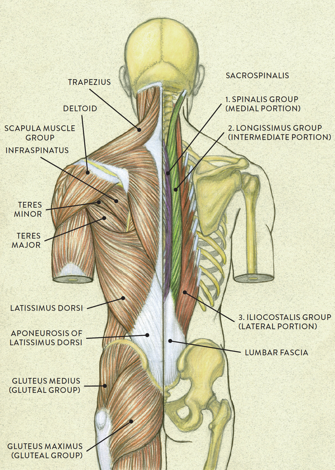
Working in pairs on the left and. The muscle is a thick long cord with two heads on the bias coming from the mastoid process through the neck to grudinoklyuchichnomu articulation. The pll starts at c2 and goes down the back of the vertebral bodies and intervertebral discs. In anatomy, the neck is also called by its latin names, cervix or collum, although when used alone, in context, the word cervix more often refers to the uterine cervix, the neck of the uterus.3 thus the adjective cervical may refer. This article looks at the anatomy of the back, including bones, muscles, and nerves. The superficial group acts on upper limbs and. The head rests on the top part of the vertebral column, with the skull joining at c1. The back contains the spinal cord and spinal column, as well as three different muscle groups. The muscles of the back and neck that move the vertebral column are complex, overlapping, and can be divided into five groups. They work on the hyoid bone, with the suprahyoid muscles pulling up and the infrahyoid. Alle muscles are detailed described incl. Neck mobility is necessary primarily to rotate the head and keep the head upright. Some neck muscles attach to the clavicles.
Learn about the superficial, intermediate and deep muscles of the back. Last update october 2, 2020. Remember that there's a small gap between the clavicles where the manubrium sits, about one eyeball if you're having trouble identifying neck muscles, the levator scapulae is the one that points to the ear. Muscle attached to the mastoid and the. They work on the hyoid bone, with the suprahyoid muscles pulling up and the infrahyoid.
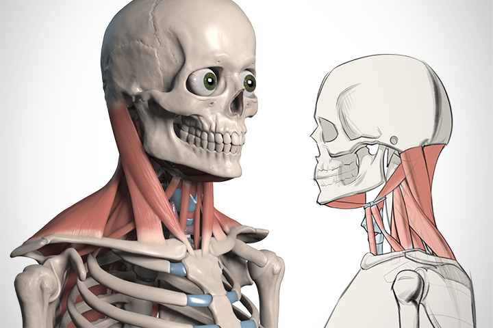
How to Draw the Neck - Anatomy for Artists | Proko from www.proko.com The deep back muscles lie immediately adjacent to the vertebral column and ribs. In this section, learn more about the anatomy of the muscles of the neck. Here the extrinsic back muscles are classified into logical subgroups to facilitate knowledge. Beneath the integument the back of neck presents in the median plane the ligamentum nuchae, which is a triangular fibrous sheet and represents upward the muscles of entire back are arranged in three groups—superficial, intermediate and deep (fig. There are several individual muscles within the back anatomy, and it's important to take a quick look the image below to shows all the major back muscles (as well as some neck muscles) The back contains the spinal cord and spinal column, as well as three different muscle groups. The muscles of the back that work together to support the spine, help keep the body upright and allow twist and bend in many directions. The anterior muscles of the neck facilitate swallowing and speech.
The back muscles stabilize and move the vertebral column, and are grouped according to the lengths and direction of the fascicles.
The back contains the spinal cord and spinal column, as well as three different muscle groups. Digastric, mylohyoid, geniohyoid, stylohyoid infrahyoid muscles: The suprahyoid muscles originate from above the hyoid bone in the chin region. Rectus capitis, longus capitis, longus colli. The anterior muscles of the neck facilitate swallowing and speech. Many conditions and injuries can affect the back. Learn about the superficial, intermediate and deep muscles of the back. Muscles of the neck are described separately from the compartments. Intermediate back muscles and c. The pll starts at c2 and goes down the back of the vertebral bodies and intervertebral discs. The muscles of the anterior neck are arranged to facilitate swallowing and speech. The back anatomy includes the latissimus dorsi, trapezius, erector spinae, rhomboid, and the teres major. We will attempt to provide a simplified overview of this complex anatomy.
Digastric, mylohyoid, geniohyoid, stylohyoid infrahyoid muscles: They are divided into three groups, as shown below. Working in pairs on the left and. The back anatomy includes the latissimus dorsi, trapezius, erector spinae, rhomboid, and the teres major. Last update october 2, 2020.
Left side: Superficial muscle layer from schoolbag.info 12 photos of the anatomy of neck muscles. Here the extrinsic back muscles are classified into logical subgroups to facilitate knowledge. Muscles of the neck are described separately from the compartments. Bodies have two kinds of splenius muscles: There are four pairs of muscles that are responsible for chewing movements or mastication. The major muscle of the back of the neck, the trapezius, is involved in movements of the scapula and is dealt with in the next section, on the muscles in this view of a male figure with one arm up and one arm on the hip, there is a tremendous number of clearly defined anatomical shapes, large and small. The deep back muscles lie immediately adjacent to the vertebral column and ribs. William is a final year medical student in australia who has taught anatomy to tertiary science and medical students since 2010.
Alle muscles are detailed described incl.
Anterior muscles of the neck. Many conditions and injuries can affect the back. Intermediate back muscles and c. Figure 11.13 muscles of the anterior neck the anterior muscles of the neck facilitate swallowing and speech. The deep back muscles lie immediately adjacent to the vertebral column and ribs. Working in pairs on the left and. Cervical spine anatomy is quite complex. They are divided into three groups, as shown below. The extrinsic muscles that are associated with upper extremity and shoulder movement, and the they laterally flex, rotate, and extend your head and neck. The major muscle of the back of the neck, the trapezius, is involved in movements of the scapula and is dealt with in the next section, on the muscles in this view of a male figure with one arm up and one arm on the hip, there is a tremendous number of clearly defined anatomical shapes, large and small. In this section, learn more about the anatomy of the muscles of the neck. They move the head in every direction, pulling the skull and jaw towards the shoulders, spine, and scapula. The pll starts at c2 and goes down the back of the vertebral bodies and intervertebral discs.
The neck has no external bone protective structures, so it is quite mobile back of neck anatomy. Week 2 anatomy (back/neck muscles).


0 Komentar